Abstract
Age-related macular degeneration (AMD) is a leading cause of central vision loss in older adults. Early stages are often subtle, and patients may remain unaware of changes until central vision is noticeably affected. Optometrists are uniquely positioned to detect these early signs through careful fundus examination, OCT imaging, and patient-reported monitoring, such as the Amsler grid.
This article explores practical strategies for recognising early AMD, monitoring disease progression, providing patient education, and determining appropriate referral. Emphasis is placed on the optometrist’s role in preventing visual deterioration and supporting timely, collaborative care with ophthalmology services.
Learning Objectives
After reading this article, optometrists should be able to:
- Recognise the early clinical features of AMD and distinguish between dry and wet forms.
- Understand the risk factors and indicators that suggest higher likelihood of progression.
- Apply practical strategies for patient education, home monitoring, and lifestyle advice.
- Identify key thresholds for routine and urgent referral to ophthalmology.
- Appreciate the importance of collaborative care pathways between optometrists and ophthalmologists.
Introduction
AMD is increasingly prevalent in older adults and can have a significant impact on independence and quality of life. Early disease is often asymptomatic, making routine eye examinations critical for detection. Subtle macular changes, including drusen and pigmentary irregularities, may be observed long before patients report visual complaints.
Optometrists play a central role in recognising these changes, monitoring progression, and advising patients on lifestyle measures and referral pathways. Through careful assessment, patient education, and collaborative practice with ophthalmologists, optometrists can significantly influence outcomes and help preserve vision.
Epidemiology and Significance
Around 10–15% of adults over 65 show early signs of AMD, while a smaller proportion develop advanced disease. Dry AMD accounts for most cases and progresses slowly, whereas wet AMD, though less common, is associated with rapid central vision loss.
In practice, early detection allows patients to be monitored and referred before substantial visual impairment occurs. The optometrist’s vigilance is therefore key in preserving visual function and maintaining patients’ quality of life.
Risk Factors
AMD arises from a combination of non-modifiable and modifiable factors. Age is the greatest predictor, followed by family history and ethnicity. Lifestyle factors, including smoking, poor diet, and cardiovascular disease, also increase risk. Smoking is particularly influential, markedly accelerating progression.
Assessment of risk factors during routine examinations helps prioritise patients for monitoring and education. Those with multiple risk factors may require more frequent review and careful documentation of subtle retinal changes.
Pathophysiology and Clinical Features
AMD affects the macula, leading to degeneration of the retinal pigment epithelium and photoreceptors. Drusen are one of the earliest signs and vary in type:
- Hard drusen – small, well-defined, usually low risk.
- Soft drusen – larger, less distinct, associated with higher risk of progression.
Early AMD may be asymptomatic. Some patients notice difficulty reading fine print, reduced contrast sensitivity, or minor distortion in straight lines. Intermediate AMD features larger or confluent drusen and pigmentary changes, while advanced disease may present as geographic atrophy (dry AMD) or abnormal blood vessel growth (wet AMD), the latter causing rapid vision loss.
Detection and Monitoring
Optometrists are well placed to identify AMD before patients experience functional vision loss. Examination techniques include:
- Dilated fundus assessment to visualise drusen and pigmentary changes.
- Slit-lamp biomicroscopy for macular evaluation.
- OCT imaging to detect fluid, cysts, or pigment epithelial detachments.
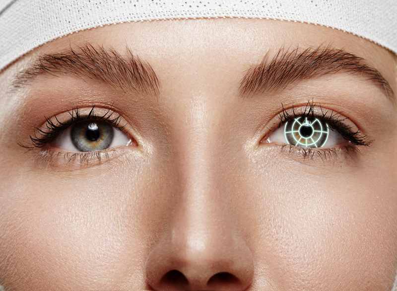
The Amsler grid remains a simple yet effective home-monitoring tool. Any reported distortion or missing lines should prompt reassessment and may indicate progression to wet AMD. Combining clinic-based evaluation with home monitoring provides a reliable picture of disease evolution and helps determine follow-up and referral intervals.
Dry vs Wet AMD
Dry AMD progresses slowly, and patients may remain largely asymptomatic for years. Monitoring involves routine evaluation, imaging, and reinforcing lifestyle measures such as diet and smoking cessation.
Wet AMD can develop rapidly, with patients reporting central distortion, blurred vision, or dark spots. OCT is critical for detecting fluid or cystic changes that require urgent referral. Early intervention with anti-VEGF therapy can substantially improve outcomes, making timely recognition and referral essential.
Patient Education and Lifestyle Guidance
Educating patients about AMD and ways to reduce risk is an integral part of care. Key points include:
- The importance of regular monitoring and home checks with an Amsler grid.
- Lifestyle measures such as smoking cessation, a diet rich in leafy greens and omega-3 fatty acids, and UV protection.
- Awareness of early warning signs such as sudden distortion or central visual changes.
Clear guidance empowers patients to act promptly and supports better visual outcomes.
Referral Considerations
Optometrists must make informed decisions regarding referral:
- Routine referral for intermediate AMD or progressive findings without acute symptoms.
- Urgent referral if patients report sudden distortion, OCT shows fluid or cysts, or central vision is affected.
Accurate documentation of retinal findings, imaging results, and patient-reported changes ensures effective communication with ophthalmology colleagues and supports timely intervention.
Reflection Questions (CPD)
- How can optometrists identify early signs of AMD during routine examinations?
- Which patient risk factors should prompt closer monitoring or earlier referral?
- How should OCT findings influence follow-up and referral decisions?
- What strategies can support patient adherence to home monitoring with the Amsler grid?
- How can lifestyle advice be integrated effectively into routine consultations?
References
- Wong WL, et al. Global prevalence of age-related macular degeneration and disease burden projection for 2020 and 2040: a systematic review and meta-analysis. Lancet Glob Health. 2014;2:e106–16.
- Cheung CMG, Arnold JJ, Holz FG, et al. Age-related macular degeneration. Lancet. 2021;398:1147–59.
- Ferris FL 3rd, Wilkinson CP, Bird A, et al. Clinical classification of age-related macular degeneration. Ophthalmology. 2013;120:844–51.
- AREDS Research Group. A randomized, placebo-controlled clinical trial of high-dose supplementation with vitamins C and E, beta carotene, and zinc for AMD. Arch Ophthalmol. 2001;119:1417–36.
- National Institute for Health and Care Excellence (NICE). Age-related macular degeneration: diagnosis and management. 2018.
- Anderson Eye Care. About Us. https://www.andersoneyecare.co.uk/about-us